Ghasem Rahimi,
Fahimeh Alizadeh ![]() ,
Alireza Khodavandi
,
Alireza Khodavandi
For correspondence:- Fahimeh Alizadeh Email: falizadeh@iauyasooj.ac.ir Tel:+987433313930
Received: 20 July 2015 Accepted: 30 January 2016 Published: 28 February 2016
Citation: Rahimi G, Alizadeh F, Khodavandi A. Mycosynthesis of silver nanoparticles from candida albicans and its antibacterial activity against Escherichia coli and Staphylococcus aureus. Trop J Pharm Res 2016; 15(2):371-375 doi: 10.4314/tjpr.v15i2.21
© 2016 The authors.
This is an Open Access article that uses a funding model which does not charge readers or their institutions for access and distributed under the terms of the Creative Commons Attribution License (http://creativecommons.org/licenses/by/4.0) and the Budapest Open Access Initiative (http://www.budapestopenaccessinitiative.org/read), which permit unrestricted use, distribution, and reproduction in any medium, provided the original work is properly credited..
Purpose: To produce and characterize silver nanoparticles using Candida albicans and evaluate its antibacterial properties.
Methods: Extracellular silver nanoparticles were biosynthesized using C. albicans. The biomass obtained from cultures of C. albicans was used to synthesize silver nanoparticles in 1.5 mM silver nitrate solution. Characterization of the biosynthesized nanoparticles was carried out using ultraviolet (UV)-visible spectrometry and scanning electron microscopy (SEM). The antibacterial properties of the nanoparticles were determined by agar disc diffusion method against Escherichia coli and Staphylococcus aureus.
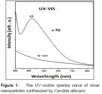
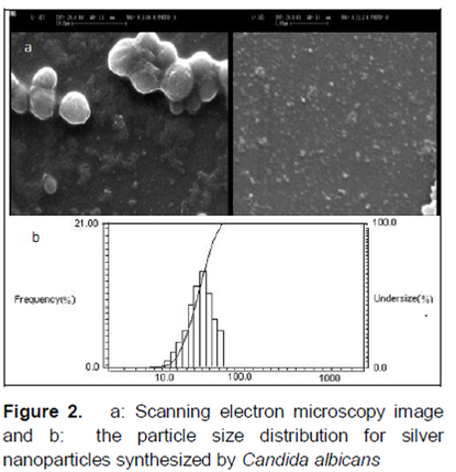
Results: Incubation of C. albicans with silver nitrate solution produced silver nanoparticles after 2 – 4 h as evidenced by change in the color of the reaction mixture. The formation of silver nanoparticles was confirmed by UV-visible spectroscopy which showed absorption peak between 300 – 800 nm, being the characteristic wavelength range of silver nanoparticles. SEM revealed the varying morphology of the nanoparticles which had a size range of 20 – 80 nm. Furthermore, the nanoparticles showed significant antimicrobial activity (p < 0.05).
Conclusion: The biosynthesized silver nanoparticles hold some promise for various industrial applications, including drug delivery.
Introduction
The use of nanotechnology in medicine is set to spread rapidly. In the last decades, the treatment of bacterial infections has become increasingly complicated due to their resistance mechanisms. This has resulted in severe debilitating and even life-threatening infections; thus this situation is driving the search for novel antibacterial agents [1].
Metal nanoparticles have proved to be effective against microorganisms. The broad-spectrum antimicrobial properties of silver encourage its widespread use [1,2]. Silver nanoparticles are of particular interest due to their distinctive physical, chemical and biological properties [3,4]. Silver nanoparticles have proved to be most effective metallic nanoparticles as they have potent antimicrobial efficacy against various pathogenic microorganisms including bacteria such as Escherichia coli, Staphylococcus aureus, Staphylococcus epidermis, Leuconostoc mesenteroides, Bacillus subtilis, Klebsiella mobilis, and Klebsiella pneumonia. Silver nanoparticles are more effective against microorganisms due to high surface area to volume ratio so that a large proportion of silver atoms are in direct contact with their environment. The antibacterial activity of silver nanoparticles against Gram negative and Gram positive bacteria have been established, but the mechanism of action of silver nanoparticles has not been completely understood [2,3,5-8].
Recently, researchers found that microorganisms have been explored as potential ecofriendly nanofactories for the synthesis of silver nanoparticles. Biosynthesis of nanoparticles has received considerable attention due to better control over size distribution of nanoparticles and the environmental toxicity [5,9,10]. The potential application of microorganisms in silver nanoparticles synthesis has been explored that can be applied to many fields and more specifically to biomedicine. Thus, significant efforts have been directed towards finding an inexpensive and environment-friendly method for nanoparticle synthesis. Several lines of evidence demonstrated the biosynthesis of extracellular silver nanoparticles utilizing many ubiquitous fungal species. Fungi especially yeast present a suitable option for large-scale green nanoproduction. The mechanism of nanoparticle production using fungi is different; fungi secrete large amounts of enzymes which are used to reduce silver ions to metal nanoparticles [3,11,12]. The mycosynthesis of silver nanoparticles was carried out from Candida albicans and their antibacterial activity against E. coli and S. aureus was evaluated.
Methods
Microorganisms
In this study 15 clinical isolates of Candida albicans were isolated and identified from immunocompromised patients. Standard strain of C. albicans ATCC 14053, Escherichia coli ATCC 39403 and Staphylococcus aureus ATCC 25923 were employed.
Biosynthesis of silver nanoparticles
The characterized isolates of C. albicans and C. albicans ATCC 14053 were plated out on Sabouraud Dextrose Agar (SDA, Merck, Germany) containing 3.5 mM silver nitrate and incubated at 37 °C for 24 h. The colonies obtained were inoculated in 250 ml Sabouraud Dextrose Broth (SDB, Merck, Germany) and incubated at 37 °C on shaking incubator at 200 rpm for 24 h. After incubation the cells were separated by centrifugation at 6,000 rpm for 15 min and washed 3 times with 50 mM phosphate buffer (pH 7.0). Two g of wet biomass was collected and then added in 1.5 mM 50 ml silver nitrate solution and incubated at 37 °C on shaking incubator at 250 rpm in dark for 48 h. The silver nitrate reduced to silver nanoparticles shows change in color of silver nitrate from colorless to brown was harvested by centrifugation at 6,000 rpm for 6 min. The production of silver nanoparticles were analyzed by UV–visible spectrophotometric analysis and subjected to scanning electron microscopy (SEM) [3].
Characterization of silver nanoparticles
The synthesis of silver nanoparticles was monitored by UV–visible spectroscopy (Shimadzu, UV Pharma spec 1700 with a resolution of 0.72 nm) by recording the spectra between 300 – 800 nm. The cell biomass of silver nitrate at time zero served as control. The produced silver nanoparticles were subjected to SEM (JEOL JSM 6360, Japan) and the images of nanoparticles were taken. The exact size and particle size distributions of silver nanoparticles were also determined by particle size analysis.
Antibacterial activity of silver nanoparticles
Silver nanoparticles synthesized from C. albicans were tested for antibacterial activity against pathogenic microorganisms E. coli ATCC 39403 and S. aureus ATCC 25923. The antibacterial activity of silver nanoparticles was tested by the agar disc diffusion method according to the Clinical and Laboratory Standards Institute protocol (M02 – A11) for bacteria with some slight modification. Both bacteria were cultured on Nutrient Agar (NA, Merck, Germany) and then passaged twice to ensure viability and purity.
Stock inoculum suspensions of the E. coli and S. aureus were prepared by picking colonies from 24 h cultures grown on NA at 37 °C and suspending in 5 ml of sterile saline (0.85 %) v/v NaCl. The suspension was adjusted with the aid of spectrophotometry at a wavelength of 625 nm to achieve turbidity equivalent to 0.5 McFarland standard, i.e., approximately (1 – 2) × 108 colony-forming units /ml). The inocula were spread on Mueller Hinton Agar using a sterile cotton swab. The culture plates were kept to dry at room temperature for 15 min. For the next step 0, 10, 20 and 50 µl of the sterile silver nanoparticles were impregnated on separate paper disks and put on the surface of cultured media. Tetracycline was used as positive control. Eventually the cultured plates were incubated at 37 °C for 24 h and then the diameter of zone of inhibition was measured. Three technical replicates were performed in each test and experiment was repeated at least two times [13].
Statistical analysis
Statistical analysis was done using SPSS software (version 21, SPSS Inc, Chicago, IL, USA). Normality and ANOVA were used to compare results. P < 0.05 was considered significant.
Results
Biosynthesized silver nanoparticles
Silver nanoparticles were synthesized from silver nitrate by C. albicans. The reaction started within 2 – 4 h and the color of the solution turned to yellowish brown, indicating the synthesis of silver nanoparticles by C. albicans.
Characteristics of silver nanoparticles
The formation of silver nanoparticles by C. albicans was further determined using the UV–visible spectroscopy, which was shown on the surface plasmon resonance bands. demonstrated that the silver nanoparticles peak was observed at 300 – 800 nm and the silver nanoparticles contribute to the absorption bands at 420 nm in the UV–visible spectra. UV–Vis spectra of aqueous silver nitrate solution (control) when monitored separately did not show any at 300 – 800 nm.
The SEM images and their corresponding particle size distributions of silver nanoparticles are shown in . The SEM images and their size distributions revealed that, the mean diameters and standard deviation of silver nanoparticles were about 20 – 80 nm with the different morphologies. Electron microscopy data indicate that the extracellular particles produced.
Antibacterial activity
The antibacterial activity of synthesized silver nanoparticles by cell biomass of C. albicans was tested against pathogenic microorganisms E. coli and S. aureus. The synthesized silver nanoparticles efficiently inhibited the growth of Gram negative and Gram positive bacteria. Our results demonstrated that the biosynthesized silver nanoparticles had more antimicrobial activity against S. aureus compared to E. coli. The zones of inhibition for S. aureus and E. coli were 21 ± 0.12 and 17 ± 0.01 mm, respectively.
Discussion
All natural silver has been recognized as a nontoxic, safe inorganic antimicrobial agent for centuries. The extracellular synthesis of silver nanoparticles had been reported numerous from bacteria, yeasts, fungi, and algae [4]. The data obtained demonstrate the extracellular synthesis of silver nanoparticles using C. albicans. These findings are consistent with Waghmare et al [3] observations that silver nanoparticles could be synthesized by Candida species. Candida is a genus of yeasts and replicate quickly, therefore are easy to manipulate genetically to make them produce more desired substances for synthesis. C. albicans is harmless commensals or endosymbiosis of hosts including humans; however, when mucosal barriers are disrupted or the immune system is compromised they can invade and cause disease [14]. Moreover, Di Giacomo et al [15] used growing cells of C. albicans in combination with carbon nanotubes to produce stable electrically conductive bio-nano-composite tissue materials that have been used as temperature sensing elements.
The appearance of brown color from colorless cell biomass of C. albicans suggested the formation of silver nanoparticles [3]. This color arises due to the surface plasmon resonance phenomenon, which monitored through the UV–visible spectrum at 420 nm, indicating formation of silver nanoparticles [3,16].
The SEM images of the particles thus obtained show different particle morphology with the size range of 20 – 80 nm. Evidence demonstrates that the morphology of nanoparticles is highly variable, and includes spherical, rod-like, decahedral, triangular, and platelet shapes. This could be due to correlation of the absorption spectrum for individual silver nanoparticles [17].
Antibacterial activity of synthesized silver nanoparticles was found to be inhibitory to both Gram negative and Gram positive bacteria. Interestingly, the synthesized silver nanoparticles have shown more antimicrobial activity against Gram positive bacteria than the Gram negative bacteria. The exact mechanism of interaction and penetration of silver nanoparticles inside the cell and their antibacterial effect remains to be identified [3,4,16,17]. Scientific evidence shows that smaller silver nanoparticles having a high definite surface area available for interaction would have more antibacterial activity than larger silver nanoparticles. Moreover, it is possible that silver nanoparticles not only interact with the surface of the membrane, but can also penetrate inside the bacteria. Furthermore, the electrostatic attraction between positively charged nanoparticles and negatively charged bacterial cell membranes could be responsible for the interactions between nanoparticles and bacterial cells [4,16,17].
The antibacterial effects of silver nanoparticles may be associated with the characteristics of certain bacterial species [4,16,17]. Our result showed the inhibition zone diameters of S. aureus were longer than those of E. coli. This implies that biosynthesized silver nanoparticles by C. albicans had better antibacterial activity against Gram positive bacteria than Gram negative bacteria, which could probably be explained by their biochemical and physiological properties [18].
The hypothesized mechanisms of antibacterial activity of silver nanoparticles, mostly relate to disturbance of the integrity of the cell membrane and depletion of the levels of intracellular ATP, ROS production, and cellular uptake of Ag+ ions or nanoparticles, due to membrane poration, with consequent suppression of respiratory enzymes and electron transport components and through interference with DNA functions [8,19,20]. Nada et al [5] demonstrated the antibacterial activity of silver nanoparticles synthesized from S. aureus against human pathogenic bacteria methicillin resistant S. epidermidis and Streptococcus pyogenes and against Salmonella typhi and K. pneumoniae. Similarly, El-Shanshoury et al [9] reported antibacterial activity of silver nanoparticles synthesized from Escherichia coli, Bacillus subtilis, and S. thermophilus against E. coli, B. subtilis, S. typhimurium, K. pneumoniae, S. aureus, Pseudomonas aeruginosa and C. albicans.
The molecular mechanisms of action of silver nanoparticles were investigated using E. coli as a model system by proteomic approach. Parallel proteomic investigations resulted in up-regulation in the expression of a panel of envelope and heat shock proteins, which are direct evidence of dissipation of the outer membrane of bacteria and deplete the levels of intracellular ATP [21].
Conclusion
The findings of this study demonstrate the simple, safe, cost-effective and ecofriendly preparation of silver nanoparticles using yeast C. albicans. The biosynthesized nanoparticles showed potential antibacterial activity against Gram-negative and Gram-positive bacteria. Thus, application of biosynthesized silver nanoparticles may lead to the development of suitable pharmaceutical and other industrial products.
Declarations
Acknowledgement
References
Archives


News Updates